Hypertrophic Cardiomyopathy (HCM) & Hypertrophic Obstructive Cardiomyopathy (HOCM)
Hypertrophic cardiomyopathy (HCM): from pathophysiology to echocardiography
Hypertrophic cardiomyopathy is a genetic disorder that causes left ventricular hypertrophy under normal loading conditions. Hypertrophic cardiomyopathy should not be confused with hypertrophy caused by increased loading conditions. Increased ventricular load is mostly caused by systemic hypertension or aortic stenosis. In hypertension, the increased systemic resistance makes it more difficult for the ventricle to eject blood into the aorta during systole. In aortic stenosis, there is increased resistance in the aortic valve itself, due to the reduced area of the valvular orifice. Both aortic stenosis and hypertension result in increased ventricular load, which the ventricle counteracts by developing hypertrophy.
Hypertrophic cardiomyopathy implies left ventricular hypertrophy under normal loading conditions.
Recommended reading
- Ventricular Pressure-Volume Relationship: Preload, Afterload, Stroke Volume, Wall Stress & Frank-Starling's law
- Myocardial Mechanics
It is fundamental to distinguish hypertrophic cardiomyopathy from hypertrophy caused by increased loading conditions. The latter is far more common and the conditions may coexist. A significant percentage of the population has hypertension, and aortic stenosis is also more common than hypertrophic cardiomyopathy (especially among elderly). Patient characteristics and the degree of hypertrophy can be used to distinguish hypertrophic cardiomyopathy from hypertrophy caused by loading conditions. In the presence of increased loading conditions, one should suspect hypertrophic cardiomyopathy if the degree of hypertrophy is disproportional to the load (i.e., if hypertrophy is more pronounced than the load could reasonably explain). The probability of hypertrophic cardiomyopathy is inversely related to age, such that the younger the patient presenting with hypertrophy, the more likely a genetic etiology.
The genetic mechanisms underlying hypertrophic cardiomyopathy are complicated and some gene variants may only cause hypertrophy under certain loading conditions (i.e in the presence of increased load). Thus, some cases of hypertrophic cardiomyopathy may be the result of a disproportionate response to increased ventricular loading.
The presence of systemic hypertension or aortic stenosis does not rule out hypertrophic cardiomyopathy.
Epidemiological aspects of hypertrophic cardiomyopathy (HCM)
Hypertrophic cardiomyopathy is equally common among men and women. The prevalence in a Western population is approximately 0.2%. Hypertrophic cardiomyopathy is one of the most common causes of cardiac arrest and sudden cardiac death (SCD) among young individuals. Among athletes, hypertrophic cardiomyopathy is the most common cause of sudden cardiac death. Therefore, current screening recommendations for athletes emphasize on measures to detect hypertrophic cardiomyopathy.
Hypertrophic cardiomyopathy is the most common cause of sudden cardiac death among athletes, and one of the most common causes of sudden cardiac death among young individuals.
Echocardiography in hypertrophic cardiomyopathy (HCM)
The hypertrophy is generally asymmetric, i.e its distribution in the left ventricular myocardium varies. Septal hypertrophy, apical hypertrophy and hypertrophy of the left ventricular free wall are common. General hypertrophy is less common.
Hypertrophic cardiomyopathy causes concentric hypertrophy
Hypertrophic cardiomyopathy causes concentric hypertrophy, which means that the generated myocardium allocates space in the ventricular cavity. In concentric hypertrophy, left ventricular volume is reduced, which means that the ejection fraction (EF) must increase to produce sufficient stroke volumes (Figure 1). Although the ventricular volume is reduced by concentric hypertrophy, it may still be normal when compared to reference values.
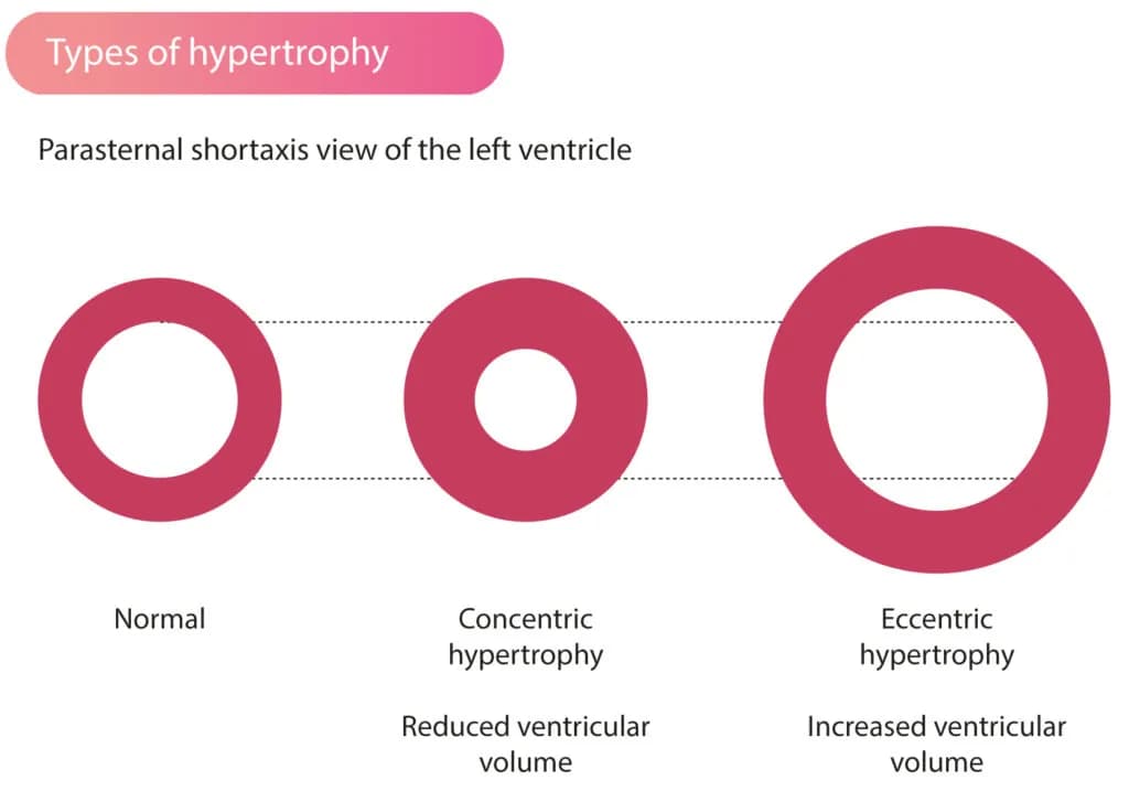
Figure 1. Types of left ventricular hypertrophy. Left ventricular volume is reduced in concentric hypertrophy. Eccentric hypertrophy results in increased ventricular volume.
The opposite of concentric hypertrophy is eccentric hypertrophy, which is common among athletes. Eccentric hypertrophy is characterized by hypertrophy of the outer myocardial layers, which does not reduce left ventricular volume. Athletes typically exhibit increased ventricular volume and slightly reduced ejection fraction. The athlete's heart is capable of maintaining cardiac output at lower ejection fractions due to the fact that they generate large stroke volumes.
Definition of hypertrophic cardiomyopathy
To diagnose hypertrophic cardiomyopathy, the following two measurements are made in the parasternal long-axis view (PLAX) or parasternal short-axis view (PSAX):
- Septal thickness
- Inferolateral wall thickness
If either exceeds 15 mm, there is hypertrophy. If the hypertrophy is not explained adequately by hypertension or aortic stenosis, hypertrophic cardiomyopathy is likely.
Athletes often display pronounced physiological hypertrophy, which can be difficult to differentiate from cardiomyopathy. Likewise, storage disorders and mitochondrial diseases can cause wall thickening, which may be difficult to differentiate from hypertrophic cardiomyopathy. The following features can be used to distinguish cardiomyopathy from the differential diagnoses:
- A hyperdynamic left ventricle suggests cardiomyopathy.
- Severe septal hypertrophy suggests cardiomyopathy.
- Obstruction in LVOT suggests cardiomyopathy.
- A small left ventricle suggests cardiomyopathy.
Table 1 presents a comprehensive list of conditions that may mimic HCM/HOCM (adapted from Marian et al [1]).
Table 1. Phenocopy Conditions for Hypertrophic Cardiomyopathy
| Phenotype | Phenotypic Clue |
|---|---|
| AMPK-mediated glycogen storage | Normal or reduced left ventricular systolic function, pre-excitation pattern |
| Pompe disease | Autosomal recessive, multiorgan disease, pre-excitation pattern |
| Anderson–Fabry disease | X-linked, multisystem also involving skin, kidney, and peripheral nerves |
| Danon disease | X-linked dominant, proximal muscle weakness, intellectual disability, short PR on ECG, elevated CK levels |
| Amyloidosis | Low QRS voltage, other organ involvement, subendothelial LGE |
| Kearns–Sayre syndrome | Multisystem disease |
| Friedreich ataxia | Autosomal recessive, neurodegeneration |
| Myotonic dystrophy | Myotonia, muscular dystrophy, cataract, and frontal baldness |
| Noonan/LEOPARD syndromes (rasopathies) | Congenital heart defects, lentigines, Café-au-lait spots |
| Neimann–Pick disease | Autosomal recessive neurodegenerative disease |
| Refsum disease | Retinitis pigmentosa, peripheral neuropathy, and ataxia |
| Deafness | Autosomal dominant deafness |
CK = creatine kinase; LGE late gadolinium enhancement.
Hypertrophic obstructive cardiomyopathy (HOCM)
In hypertrophic cardiomyopathy, it is important to clarify whether the hypertrophy causes a narrowing of the left ventricular outflow tract (LVOT). Approximately 65% of patients with hypertrophic cardiomyopathy have obstruction in LVOT, a condition referred to as hypertrophic obstructive cardiomyopathy (HOCM).
HOCM with systolic anterior motion (SAM)
The obstruction in LVOT is caused by septal hypertrophy. When the septum bulges into the LVOT, hemodynamics change in the outflow tract, which leads to the anterior leaflet of the mitral valve being sucked into the LVOT. As a result, the outflow tract is obstructed. The motion of the anterior leaflet of the mitral valve is called systolic anterior motion (SAM). Thus, obstruction of the LVOT is due to hypertrophy of the septum and subsequent SAM (Figure 2).
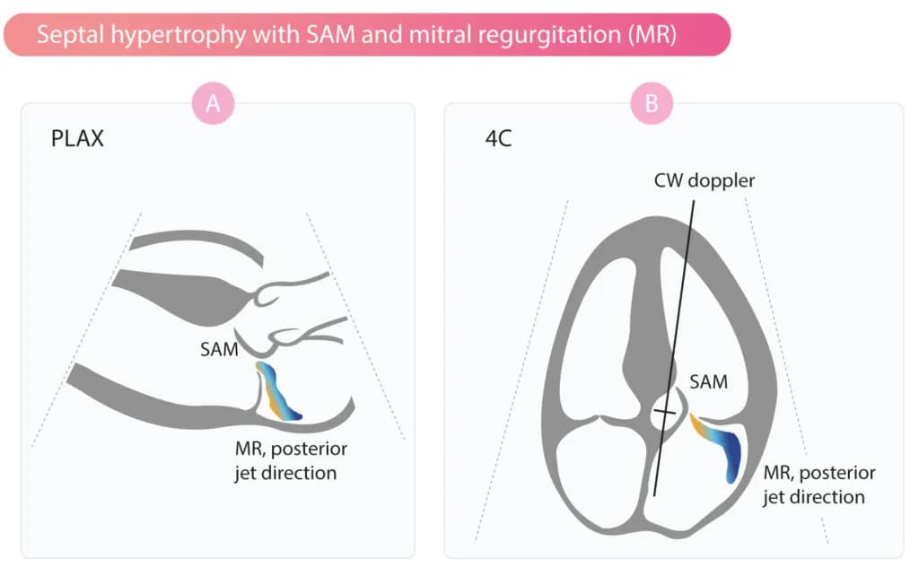
Figure 2. Systolic anterior motion (SAM) of the anterior leaflet of the mitral valve causes obstruction of the LVOT. SAM is typically accompanied by mitral regurgitation (MR), with posteriorly directed jet. CW = continuous wave doppler.
If SAM is pronounced, the anterior leaflet may touch the septum during systole. Subsequently, a pronounced obstruction can lead to closure or flutter of the aortic valve during systole.
Mitral regurgitation is a byproduct of SAM (Figure 2).
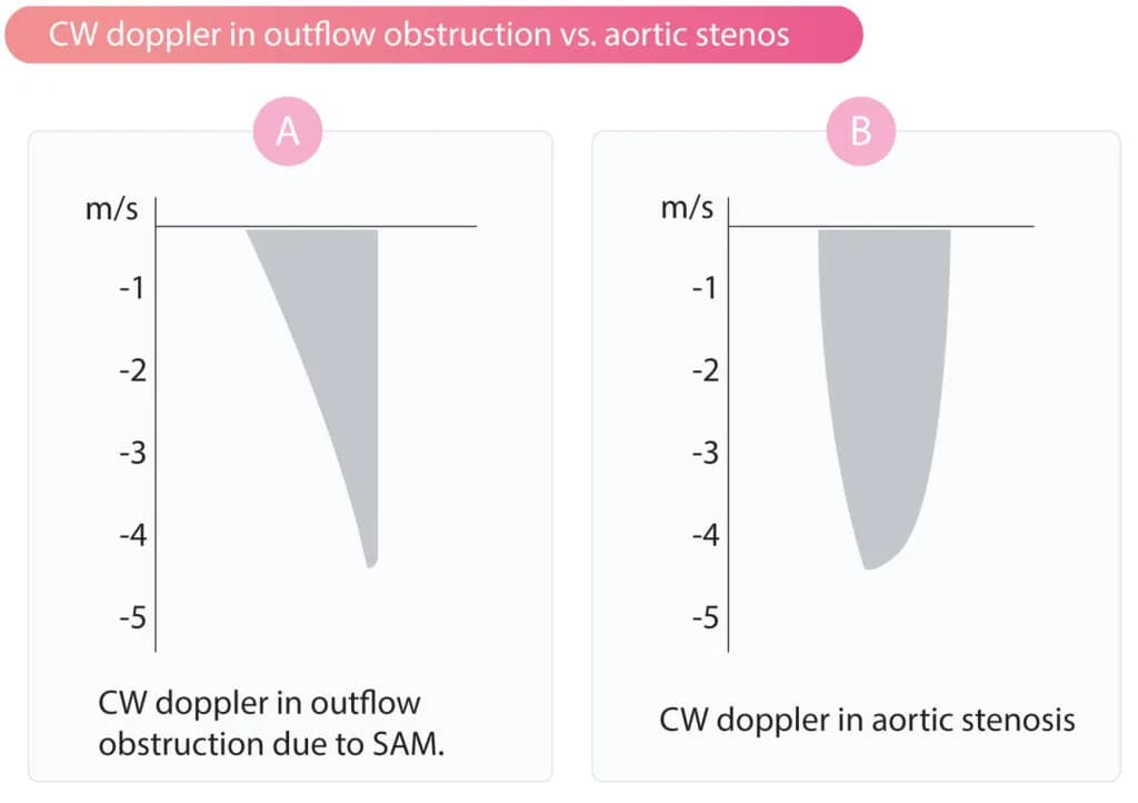
Figure 3. Continuous wave (CW) doppler in the LVOT in (A) obstruction due to SAM and septal hypertrophy and (B) aortic stenosis.
Continuous wave (CW) doppler is used to detect obstruction in the LVOT (Figures 2 & 3). The spectral curve is characterized by a slow acceleration, which distinguishes it from the Doppler signal in aortic stenosis (Figure 3).
Note that SAM typically causes the mitral valve regurgitation jet to involve the LVOT. It is important to place the Doppler cursor correctly in the LVOT in order to avoid unintentional recording of the mitral valve regurgitation jet. Video 1 shows HOCM with SAM.
Video 1. HOCM with SAM.
Obstruction in the LVOT is affected by left ventricular filling. The less the filling, the more pronounced the obstruction. This implies that hypovolemia and tachycardia (both lead to diminished ventricular filling) cause increased obstruction in the LVOT. Valsalva maneuver also reduces left ventricular filling (obstruction in LVOT can be provoked by performing Valsalva maneuver).
SAM causes mitral regurgitation (MR)
As mentioned above, hypertrophic cardiomyopathy with SAM is generally accompanied by mitral valve regurgitation (MR) with a posteriorly directed jet.
Apical and midventricular hypertrophy
In midventricular hypertrophy, obstruction may be observed midventricularly, which is detected using continuous wave (CW) Doppler (Figure 4A). In apical hypertrophy, thickened myocardium is seen in the apex. This gives the cavity a pointed appearance, as demonstrated in Figure 4B. Patients with apical hypertrophic cardiomyopathy exhibit T-wave inversion in the precordial leads (V1-V6) on ECG.
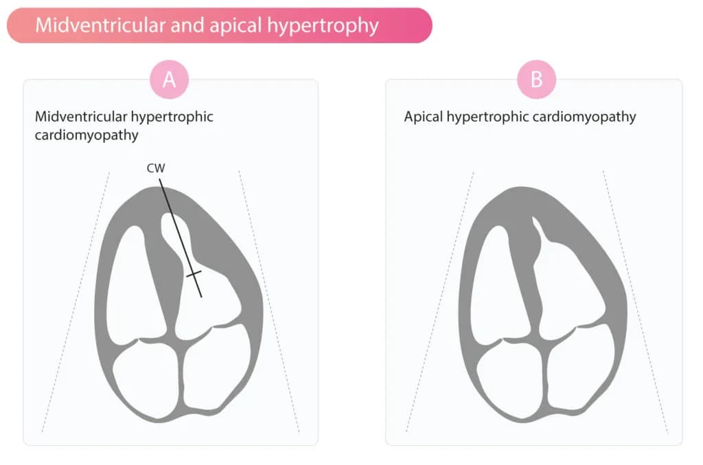
Figure 4. Apical and midventricular hypertrophic cardiomyopathy.
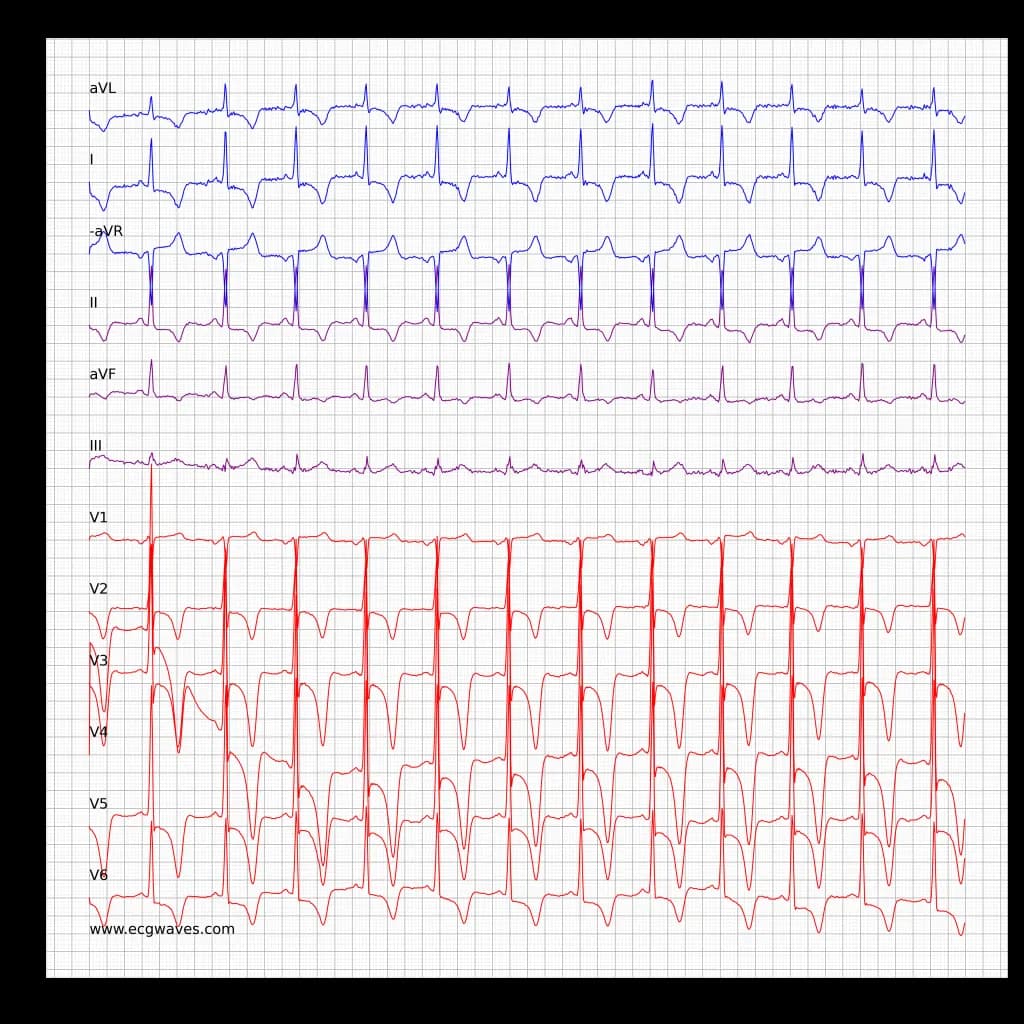
ECG 1. ECG in hypertrophic obstructive cardiomyopathy (HCM, HOCM)
Diastolic function in hypertrophic cardiomyopathy
Hypertrophic cardiomyopathy leads to impaired diastolic function, i.e the relaxation of the left ventricle is impaired, resulting in prolonged deceleration time (DT) and reduced E/A ratio. The deceleration time is prolonged because it takes longer to equalize the pressure difference between the left atrium and the ventricle. This is explained by the fact that left ventricular compliance is reduced in hypertrophic cardiomyopathy.
Sudden Cardiac Death (SCD) in hypertrophic cardiomyopathy
Hypertrophic cardiomyopathy is one of the most common causes of sudden cardiac arrest among young people. Cardiac arrest can strike any individual with hypertrophic cardiomyopathy. It should be noted, however, that the incidence of sudden cardiac arrest is very low among people with HCM/HOCM.
Patients with hypertrophic cardiomyopathy who have experienced circulatory arrest or malignant ventricular arrhythmias are unlikely to benefit from beta-blockers or antiarrhythmic drugs. The most effective treatment is an ICD (Implantable Cardioverter Defibrillator). Table 2 shows risk factors for cardiac arrest in hypertrophic cardiomyopathy.
Table 2. Risk factors for sudden cardiac arrest in cardiomyopathy
| Known riskfactors |
| Previous cardiac arrest (“aborted SCD”) |
| Family history of sudden cardiac arrest |
| Previous syncope |
| History of ventricular tachycardia |
| Severe hypertrophy |
| Probable riskfactors |
| LVOT obstruction |
| Abnormal blood pressure reaction during exercise |
| Early onset of symptoms |
Pitfalls
SAM also occurs in individuals who do not have HOCM. Individuals who have left ventricular hypertrophy may develop SAM in the setting of hypovolemia.
Below follows supplementary material intended for readers interested in the genes causing HCM. Refer to Marian et al for details (1).
Established causal genes for HCM
Established causal gene HCM (large families)
| Gene | Protein | Function |
|---|---|---|
| MYH7 | β-Myosin heavy chain | ATPase activity, force generation |
| MYBPC3 | Myosin-binding protein C | Cardiac contraction |
| TNNT2 | Cardiac troponin T | Regulator of actomyosin interaction |
| TNNI3 | Cardiac troponin I | Inhibitor of actomyosin interaction |
| TPM1 | α-Tropomyosin | Places the troponin complex on cardiac actin |
| ACTC1 | Cardiac α-actin | Actomyosin interaction |
| MYL2 | Regulatory myosin light chain | Myosin heavy chain 7–binding protein |
| MYL3 | Essential myosin light chain | Myosin heavy chain 7–binding protein |
| CSRP3 | Cysteine- and glycine-rich protein 3 | Muscle LIM protein (MLP), a Z disk protein |
Likely causal genes for HCM (small families)
| Gene | Protein | Function |
|---|---|---|
| FHL1 | Four-and-a-half LIM domains 1 | Muscle development and hypertrophy |
| MYOZ2 | Myozenin 2 (calsarcin 1) | Z disk protein |
| PLN | Phospholamban | Regulator of sarcoplasmic reticulum calcium |
| TCAP | Tcap (telethonin) | Titin capping protein |
| TRIM63 | Muscle ring finger protein 1 | E3 ligase of proteasome ubiquitin system |
| TTN | Titin | Sarcomere function |
Genes associated with HCM (small families and sporadic cases)
| ACTN2 | Actinin, α2 | Z disk protein |
| ANKRD1 | Ankyrin repeat domain 1 | A negative regulator of cardiac genes |
| CASQ2 | Calsequestrin 2 | Calcium-binding protein |
| CAV3 | Caveolin 3 | A caveolae protein |
| JPH2 | Junctophilin-2 | Intracellular calcium signaling |
| LDB3 | Lim domain binding 3 | Z disk protein |
| MYH6 | Myosin heavy chain α | Sarcomere protein expressed at low levels in the adult human heart |
| MYLK2 | Myosin light chain kinase 2 | Phosphorylate myosin light chain 2 |
| NEXN | Nexilin | Z disc protein |
| TNNC1 | Cardiac troponin C | Calcium-sensitive regulator of myofilament function |
| VCL | Vinculin | Z disk protein |
References
Echocardiography in cardiomyopathies: an overview
Cardiomyopathy implies that the myocardium is structurally and functionally altered, and the pathological changes are not explained by coronary heart…
Dilated Cardiomyopathy (DCM): Definition, Types, Diagnostics & Treatment
Dilated cardiomyopathy DCM is defined as dilation of one or both ventricles. Dilation of the left ventricle is virtually always accompanied by impaired left…