Biventricular hypertrophy ECG and clinical characteristics
Biventricular hypertrophy Simultaneous hypertrophy of the left and right ventricle is rather common. The ECG has low sensitivity with respect to detecting biventricular…
Biventricular hypertrophy
Simultaneous hypertrophy of the left and right ventricle is rather common. The ECG has low sensitivity with respect to detecting biventricular hypertrophy. This is because the strong and oppositely directed forces tend to cancel each other out. However, the ECG may show features characteristic of biventricular hypertrophy.
Biventricular hypertrophy ECG
In case the ECG or other examinations suggest left ventricular hypertrophy (LVH), one should suspect concomitant right ventricular hypertrophy (RVH) if the following ECG criteria are presented:
- Right axis deviation (>90°) - This never occurs in LVH.
- Deep S-wave in V5 or V6 (>6 mm)
- Large RS complexes in multiple leads
- P pulmonale

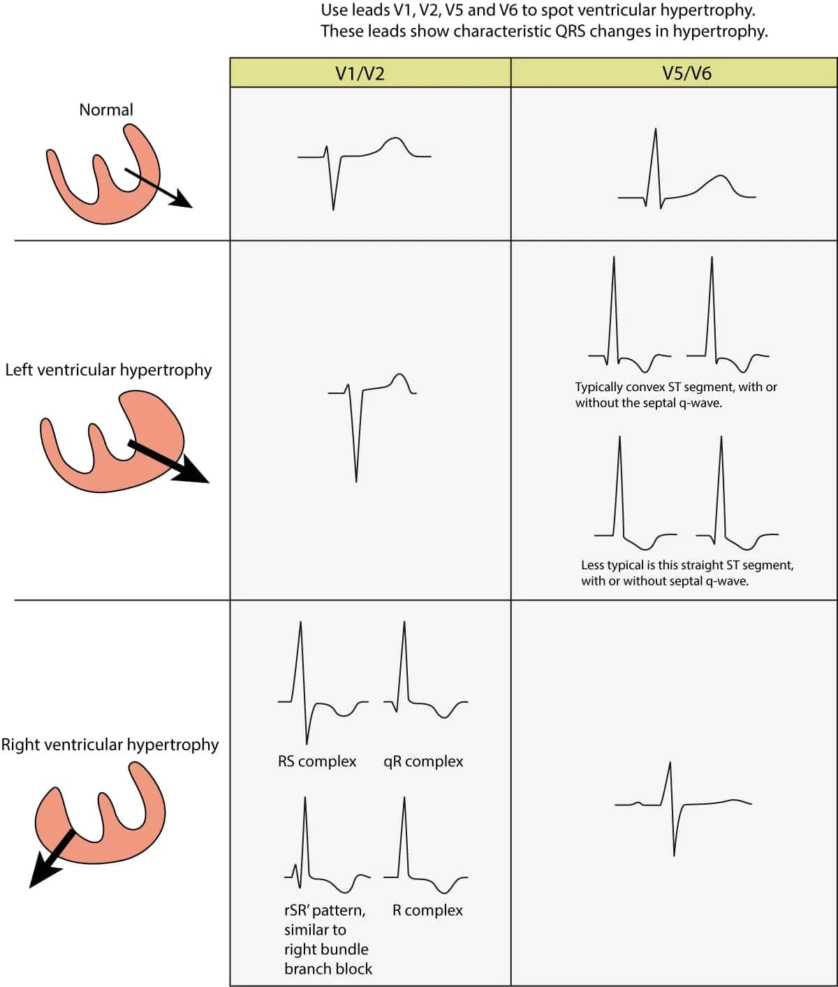
Figure 1. ECG changes seen in left ventricular hypertrophy (LVH) and right ventricular hypertrophy (RVH). The electrical vector of the left ventricle is enhanced in LVH, which results in large R-waves in left sided leads (V5, V6, aVL and I) and deep S-waves in right sided chest leads (V1, V2). Right ventricular hypertrophy causes large R-waves in right sided chest leads and deeper S-waves in left sided leads.
Right ventricular hypertrophy (RVH): ECG criteria & clinical characteristics
ECG and clinical characteristics in right ventricular hypertrophy (RVH) Figure 1. ECG changes seen in left ventricular hypertrophy (LVH) and right ventricular hypertrophy (RVH).…
Left atrial enlargement (P mitrale) & right atrial enlargement (P pulmonale) on ECG
Atrial enlargement: left atrial enlargement (P mitrale) and right atrial enlargement (P pulmonale) The atria may become dilated and/or hypertrophic during pathological…