A new ECG biomarker for sudden cardiac death
Published 2026-06-28
A deep-learning ECG biomarker for sudden cardiac death: what the new Nature study shows
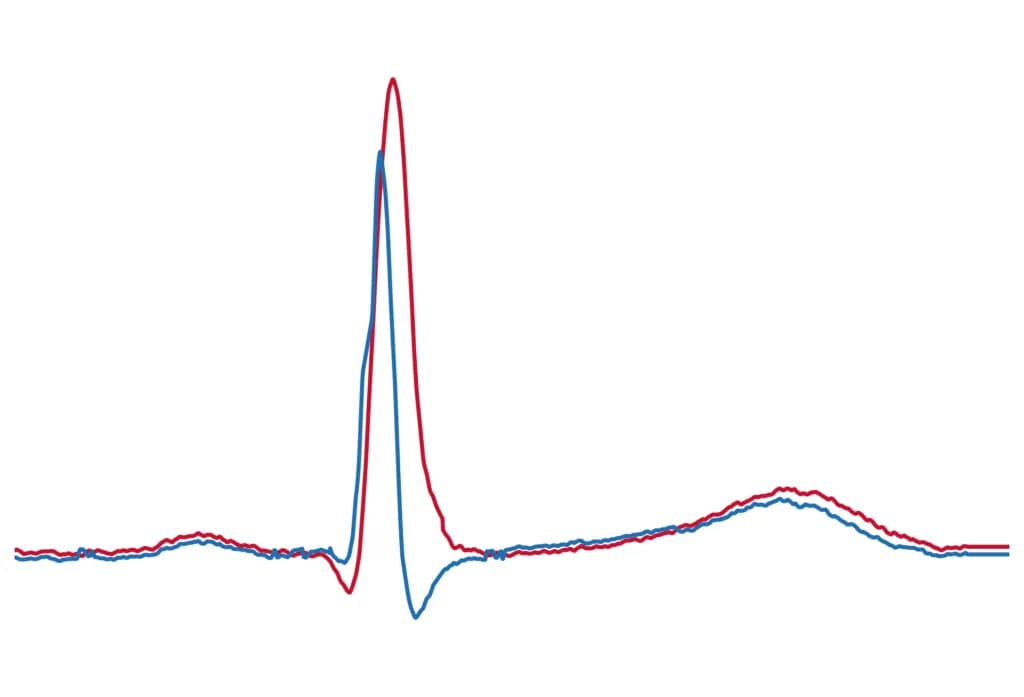
Low-risk (blue) versus high-risk (red) ECG morphs in lead aVL (adapted from Obermeyer et al1). Representative single-beat morphs for a low-risk patient (blue) and the corresponding high-risk morph (red). Beyond the difference in R-wave amplitude, note the slurred terminal portion of the R wave in the high-risk morphology, which replaces the sharp negative S wave seen in the low-risk beat — the previously undescribed biomarker identified by the deep-learning model.
Predicting sudden cardiac arrest
Sudden cardiac death (SCD) remains one of the most intractable challenges in clinical cardiology. Hundreds of thousands of arrhythmic deaths occur annually in the United States alone, yet no reliable model currently exists for the long-term prediction of sudden cardiac arrest. The constraint does not lie in therapy: implantable cardioverter-defibrillators (ICDs) are highly effective, terminating well over 99% of the ventricular arrhythmias they detect. It lies instead in patient selection. Because implantation entails appreciable cost and procedural risk, the risk-benefit balance depends almost entirely on the capacity to identify, prospectively, those individuals genuinely at risk of arrest. This predictive problem has proven persistently resistant to resolution.
Known predictors of cardiac arrest
The only risk biomarker in genuinely widespread use is left ventricular ejection fraction (LVEF), measured by echocardiography. Reduced LVEF reliably identifies a high-risk group that derives a large, trial-proven survival benefit from primary-prevention ICDs. Its dominance, though, owes as much to pragmatics as to discrimination: echocardiography is ubiquitous, standardized, and cheap to interpret.
The limitation, familiar to every electrophysiologist, is that LVEF is both insensitive and non-specific for SCD. Most people who die suddenly never had an ejection fraction measured before death, and among those who did, only a minority had a reduced value — the majority of sudden cardiac deaths occur in patients with preserved LVEF, for whom no practical risk-prediction tool currently exists. At the other end, roughly two-thirds of ICDs implanted for reduced LVEF never deliver an appropriate shock, so the false-positive burden is substantial.
Alternative modalities — cardiac MRI, long-term ambulatory monitoring, electrophysiological study, PET, SPECT, genetic profiling — add information but are too costly or invasive to screen a population. The surface ECG is the obvious cheap, standardized, universally available alternative, and it has been mined for predictive features for decades. The problem has been that hand-engineered ECG markers have never matched LVEF, and extracting them depends on expert annotation that limits scale and generalizability. Deep learning offers a way around this: an ECG can be fed directly into a classifier with no human feature extraction, in principle delivering LVEF's practical advantages with better discrimination. The missing ingredient has been a training set large enough to link raw waveforms to reliable cause-of-death data.
The current study
This paper supplies that ingredient. The authors trained a convolutional deep-learning model on population-scale ECG data from a single Swedish region, linked to national death certificates and electronic health records, and asked whether it could isolate a clinically actionable high-risk group for SCD — one defined entirely from the ECG, without echocardiography. They then stress-tested the model against multiple outcome definitions and validated it, with no fine-tuning, in geographically and structurally distinct US and Taiwanese cohorts. Finally, and unusually, they paired the predictor with a generative model to visualize the waveform morphology the network had learned, converting an opaque classifier into a human-readable, hypothesis-generating biomarker.
What they studied
The work addresses three linked questions. First, can a model trained only on raw 12-lead ECGs flag a discrete population whose annual SCD risk is high enough to justify a defibrillator? Second, is that signal specifically arrhythmic — predicting the ventricular fibrillation and ventricular tachycardia (VF/VT) that actually cause sudden death — rather than a generic marker of cardiovascular mortality? Third, what is the model actually "seeing," and does it correspond to a recognizable electrophysiological substrate?
Study data
The Swedish cohort comprised all 441,614 ECGs recorded between 2010 and 2016 in Region Halland (a public regional health system serving roughly 187,677 patients), sampled at 500 Hz and retrieved from a Philips IntelliSpace archive. Methodologically, the most important design choice is the data "lockbox": 40% of patients, with all their ECGs, were randomly partitioned off before any analysis and left untouched through model development and peer review, opened only after provisional acceptance. The model was initially trained and validated on the other 60% (262,554 ECGs from 75,157 patients) and then applied without modification to the lockbox (119,541 ECGs with one-year follow-up from 35,885 patients under 80; 113,072 ECGs in the defibrillator-naive subset). This is a far stronger guard against overfitting and optimistic reporting than the usual single train/test split, and reassuringly, performance improved rather than degraded when moving to the held-out data.
The architecture itself is a 64-layer ResNet (32 residual blocks), trained in a multitask setup that simultaneously predicted SCD over several horizons, SCD versus other causes of death among decedents, and reduced LVEF among those with echocardiography — the last giving the network an LVEF-relevant signal without requiring LVEF at inference.
External validation used two deliberately heterogeneous datasets. The US cohort (Sharp HealthCare, via Dandelion Health) contributed 251,858 ECGs from 139,613 patients under 80, recorded in 2021–2022 on GE MUSE equipment — a different vendor, format, and era from the Swedish training data. The Taiwanese dataset (National Taiwan University Hospital, via Nightingale Open Science) is a case–control ED registry of 257 cardiac arrests and 4,011 controls under 80, with detailed adjudication that classified 96 arrests (37%) as arrhythmic in origin.
Outcomes studied
Recognizing that no single SCD definition is fully reliable, the authors triangulated across several. The primary label was death-certificate SCD — cardiac or ill-defined cause, out-of-hospital or within the first 24 hours of admission — in the year following an ECG, using standard epidemiological criteria. To counter the known specificity problems of death certificates (which can misclassify non-arrhythmic deaths from stroke or pulmonary causes), they additionally evaluated against incident VF/VT documented in health records in both Sweden and the US, and against the detailed case-adjudicated arrhythmic arrests in Taiwan. As a fourth line of evidence, they estimated potential mortality benefit by comparing high-risk patients who did and did not receive defibrillators.
What they discovered
In the Swedish lockbox, the model achieved an AUC of 0.872 (95% CI 0.843–0.899) for death-certificate SCD, substantially exceeding both a generic AHA/ACC 10-year risk score (0.697) and a previously validated ECG-based deep-learning cardiovascular risk model (SEER, 0.655).
Discrimination only matters clinically if it yields a usable threshold. Anchoring to the control-group event rates from six major primary-prevention defibrillator trials (median ~4.9% annual SCD — the level at which enrolment was considered ethically justified), the authors defined a preferred high-risk group of 2.2% of the sample with a 7.0% annual SCD rate (95% CI 4.9–9.5%). For comparison, the reduced-LVEF group (≤35%) represented 1.9% of the sample with a 4.6% annual rate. Critically, 86.1% of the model's high-risk patients were not flagged by reduced LVEF — this is not a rediscovery of the echocardiographic high-risk group but a largely net-new population. Even among patients with a measured and normal LVEF, for whom no risk tool currently exists, the model isolated a subgroup at 6.4% annual risk, exceeding the reduced-LVEF group. Where both biomarkers agreed, risk rose to 10.7%, indicating that LVEF carries independent information rather than being subsumed.
The arrhythmia-specificity analyses are what elevate this from "another ECG risk score." Beyond the 7.0% with SCD, an additional 3.8% per year of the high-risk group had documented VF/VT. In the US cohort — where financial incentives drive more complete VF/VT coding — the zero-shot AUC for VF/VT was 0.822, and the matched 2.2% high-risk group showed a 29.1% VF/VT incidence against a 3.8% base rate. The Taiwanese case–control data provided the cleanest test of specificity: the model distinguished adjudicated arrhythmic arrests from controls with a zero-shot AUC of 0.767, while a "placebo" AUC for non-arrhythmic arrests (pulmonary, neurological) fell to 0.582 — close to chance, and significantly worse (P < 0.001). The signal is arrhythmic, not a generic death predictor.
On preventability, observational regressions (necessarily confounded, and presented as suggestive) found that high-risk patients with a defibrillator in place died 3.62 percentage points below their predicted 6.65% rate — a 54.4% relative reduction in SCD (P < 0.001). As a face-validity check, the same framework "rediscovered" the known LVEF effect (67.5% reduction, within the 50–88% range of LVEF-enrolled trials). The ECG high-risk group also showed a 12.6-point (39.0%) reduction in all-cause mortality with defibrillators, whereas the reduced-LVEF group did not reach significance for all-cause mortality — consistent with competing-cause mortality eroding benefit in the older, sicker LVEF population.
The biomarker itself was made visible by coupling the predictor to a variational autoencoder generative model, then iteratively morphing a real low-risk beat along the predicted-risk gradient into its high-risk counterfactual. The morph reproduced familiar correlates of ischemic disease — left axis deviation consistent with left anterior fascicular block (LAFB), and poor R-wave progression. More interesting is a feature in lead aVL that, to the authors' knowledge, has not been described: a slurred terminal aspect of the R wave replacing the sharp negative S wave seen in low-risk beats. Quantified as the mean absolute first and second voltage differences from R peak to QRS end, this feature independently and significantly predicted SCD in Sweden and the US (and trended the same way in the smaller Taiwanese sample), adding signal of comparable magnitude to left axis deviation and distinct from intrinsicoid deflection, fragmented QRS, late potentials, and QRS duration. Single-lead models retained nearly the full AUC, implying a diffuse myocardial process rather than a focal one.
That diffuseness motivated a mechanistic hypothesis. A slurred, progressively orthogonal terminal vector is what one would expect if randomly distributed obstacles repeatedly split the depolarization wavefront — and blinded cMRI review found that the riskiest 10% of patients had significantly more subtle, diffuse late gadolinium enhancement throughout the left ventricle, the imaging signature of myocardial fibrosis. Fibrosis plausibly unifies the biomarker, the LAFB, and the arrhythmic risk, since the left anterior fascicle is thin and vulnerable to scattered lesions. Notably, the authors found no mention of this LGE in any of the corresponding cardiologists' reports — a reminder that subtle diffuse fibrosis is easily overlooked and frequently false-negative even on MRI relative to biopsy.
Differences and similaritites to early repolarization pattern
Early repolarization and the deep-learning–derived sudden cardiac death biomarker are both abnormalities of the terminal portion of the QRS complex, and the parallels between them are worth understanding alongside their important differences. In each, the morphological change occurs in the late, downslope region of the QRS rather than in its initial deflections, and in each the abnormality has prognostic weight for ventricular arrhythmia and sudden cardiac death. Both can be appreciated on a standard resting twelve-lead ECG without specialized acquisition, and both followed a similar historical trajectory: early repolarization was long classified as a benign normal variant before its association with idiopathic ventricular fibrillation established it as a potentially malignant entity within the J-wave syndromes, while the newer biomarker likewise identifies a high-risk waveform that had not previously been characterized.
The two diverge, however, in both morphology and mechanism. Early repolarization is defined by J-point elevation manifesting as slurring or notching at the junction of the QRS and ST segments, typically in the inferior and inferolateral leads, and reflects a transmural repolarization gradient mediated by the transient outward potassium current between epicardium and endocardium. The deep-learning biomarker, by contrast, lies within the QRS itself — a slurred terminal R wave that replaces the sharp S wave, most evident in lead aVL and accompanied by left axis deviation and poor R-wave progression — and is attributed not to a repolarization gradient but to a depolarization and conduction disturbance, specifically diffuse myocardial fibrosis that fragments and scatters the activation wavefront. A practical consequence of this distinction is that early repolarization adds a discrete deflection that tends to increase the high-frequency content of the terminal QRS, whereas the new pattern represents a smoothing of the waveform, quantified as a reduction in the first and second voltage derivatives over the same interval. In summary, the two share an anatomical locus on the electrocardiogram and a common association with sudden cardiac death, yet they represent fundamentally distinct electrophysiological processes — one a transmural repolarization phenomenon, the other a conduction abnormality rooted in structural fibrosis.
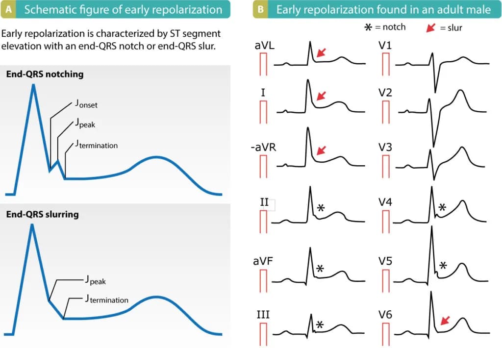
Figure 5. Early repolarization (after MacFarlane et al, 2016, Eur Hear J).
Conclusions
The practical message is that a single, cheap, already-ubiquitous test — the resting 12-lead ECG — can identify a large, previously unsuspected population at high, arrhythmia-specific, and plausibly preventable risk of sudden death, the great majority of whom are invisible to LVEF. Validation across Sweden, the US, and Taiwan, with no fine-tuning and across different vendors and outcome definitions, is unusually robust, and the lockbox design lends credibility to the headline numbers.
The appropriate caution is equally clear. The defibrillator-benefit estimates are observational and drawn from patients already selected for implantation under current norms; they cannot establish that a newly identified high-risk patient would benefit. The authors are explicit that a randomized trial in this ECG-defined population is the necessary next step — and the history of SCD prediction is littered with promising markers that failed to identify patients who actually benefit from devices. Subcutaneous and wearable defibrillator technology, however, lowers the threshold for testing such groups.
References
Blog
Articles and clinical commentary on cardiovascular medicine.
Incretins in Cardiometabolic Medicine: Leveraging GLP-1, GIP, and Glucagon Receptor Agonism for Cardiovascular Risk Reduction
Cardiovascular medicine is currently witnessing a paradigm shift. For decades, the management of type 2 diabetes mellitus (T2DM) and obesity was siloed, with clinicians focusing…