The Cardiovascular Atlas

CVD Atlas

Figure 2. Left bundle branch block (LBBB) on ECG

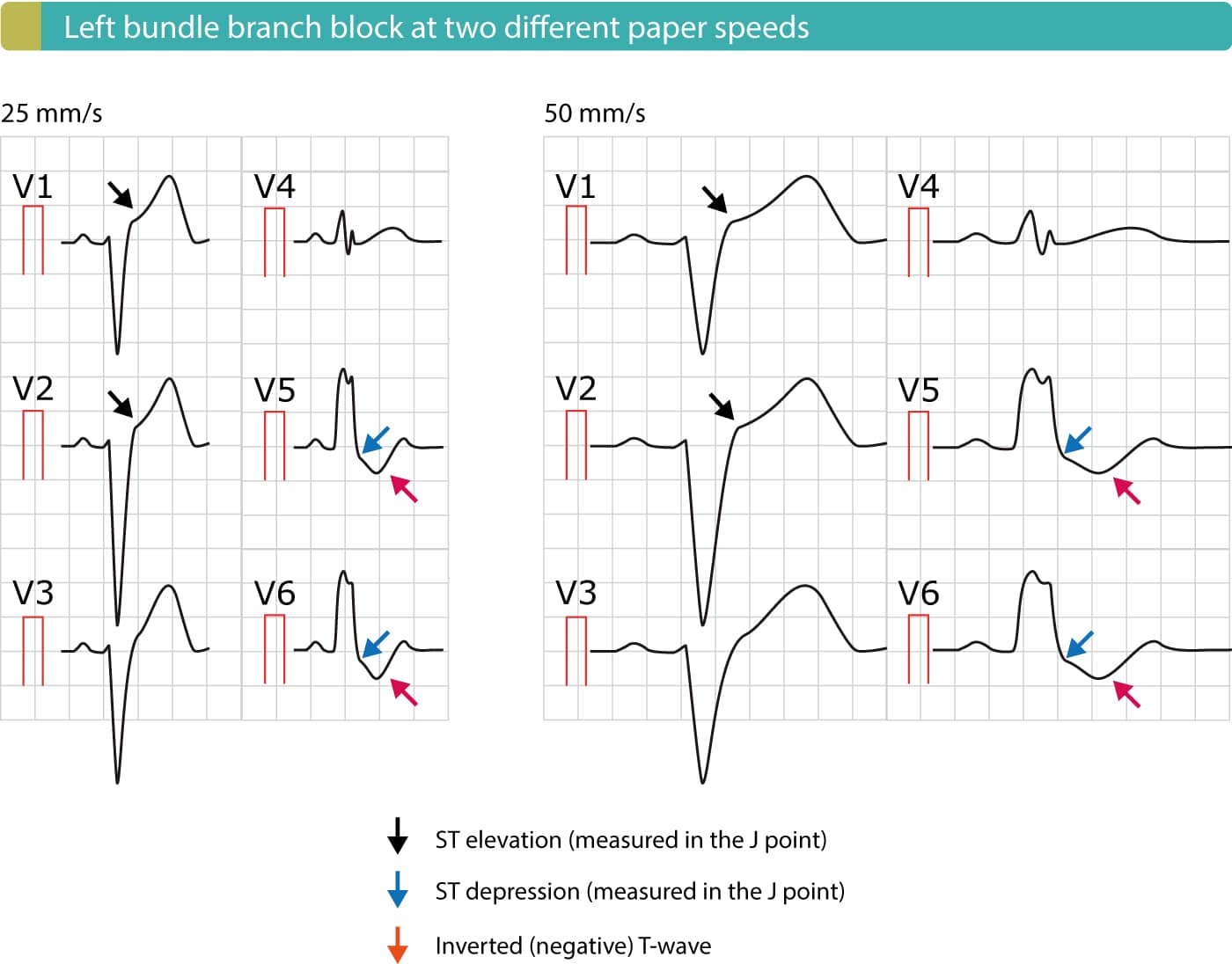
Figure 2. Left bundle branch block (LBBB) at two different paper speeds (25 mm/s and 50 mm/s). Because activation (depolarization) of the left ventricle is abnormal in the presence of LBBB, the recovery (repolarization) will also be abnormal. Abnormal depolarization manifests as an abnormal QRS complex with duration 120 ms or more. Abnormal repolarization manifests with ST-T changes, including ST elevations, ST depressions and negative T-waves.

Figure 2. Left bundle branch block (LBBB) at two paper speeds (25 mm/s and 50 mm/s). Because activation (depolarization) of the left ventricle is abnormal in the presence of LBBB, the recovery (repolarization) will also be abnormal. The abnormal depolarizatin manifests as an abnormal QRS complex with duration 120 ms or more. The abnormal repolarization manifests with ST-T changes, including ST elevations, ST depressions and negative T-waves.

CONTINUE LEARNING

Evidence-based Cardiovascular Medicine.

Peer-Reviewed.

Trusted by experts worldwide.