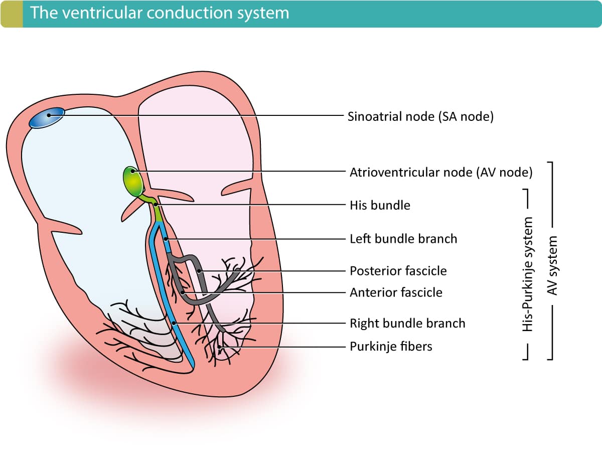
Figure 1. The electrical conduction system of the heart, with emphasis on the ventricles. The sinoatrial (SA) node and atrioventricular (AV) node are located in the atria (they are not part of the ventricular conduction system). The AV node continues in the bundle of His, which branches into the right and left bundle branch. The left bundle branch is then divided into an anterior and posterior fascicle. The right bundle branch and the fascicles then branch further into the fine Purkinje network which sprouts out to the entire myocardium. Note that the ventricular conduction system is the His-Purkinje system.