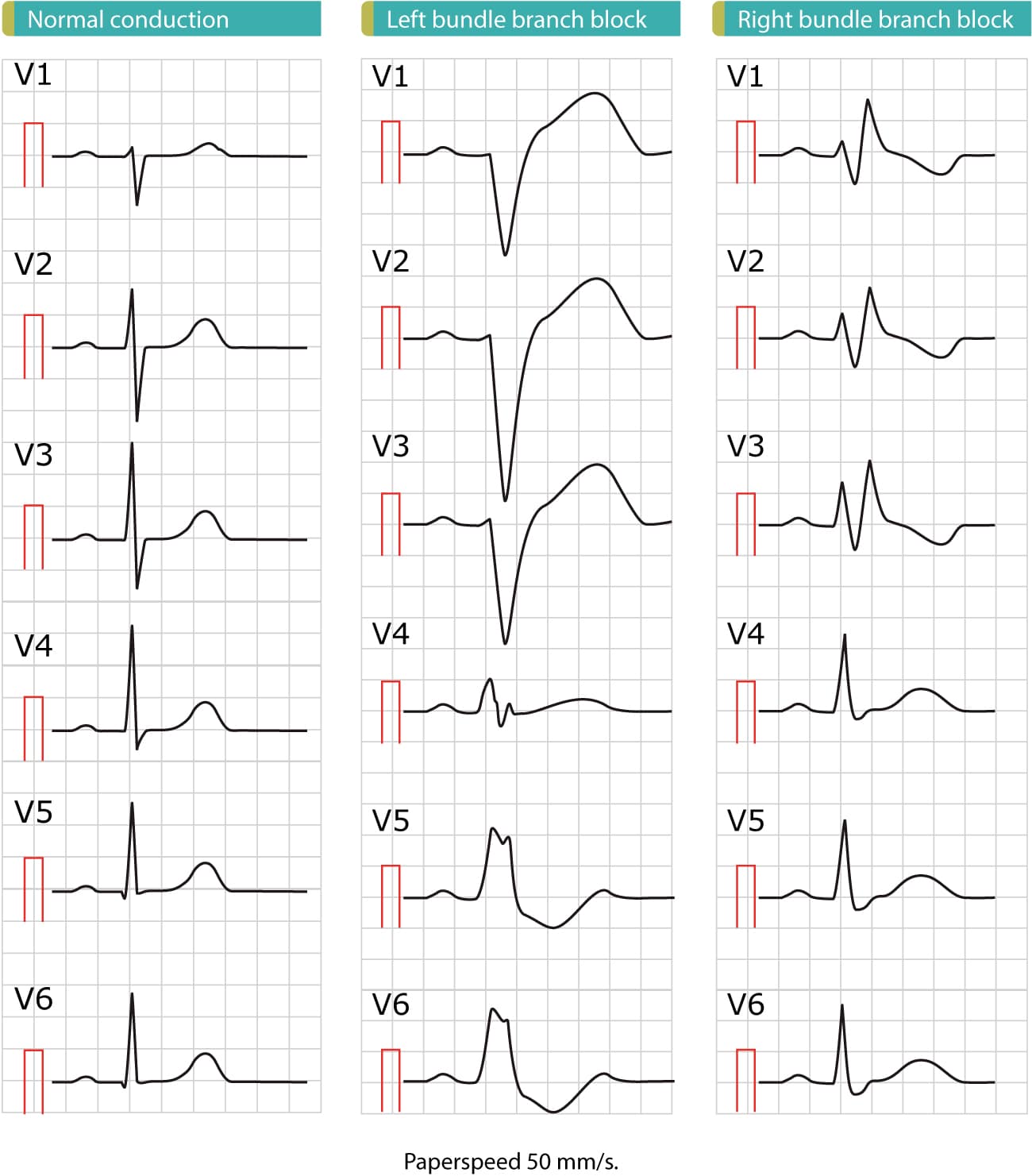
Figure 1. These ECGs show the difference between normal conduction, left bundle branch block (LBBB) and right bundle branch block (RBBB). As evident from these ECGs, the cardinal difference between normal conduction and bundle branch blocks is the QRS duration: bundle branch blocks are caused by dysfunctional bundle branches, which results in slow (and abnormal) activation of ventricular myocardium and thus prolonged QRS duration. A QRS duration of 120 ms (0.12 s) is required to diagnose bundle branch block. Also note that both left bundle branch block (LBBB) and right bundle branch block (RBBB) cause marked ST-T changes, including ST elevations, ST depressions and inverted (negative) T-waves. These ST-T changes are due to abnormal repolarization and they are expected to occur in both LBBB and RBBB. Finally, note that there are marked difference in the ECG pattern between LBBB and RBBB. These ECGs are printed at paperspeed 50 mm/s.