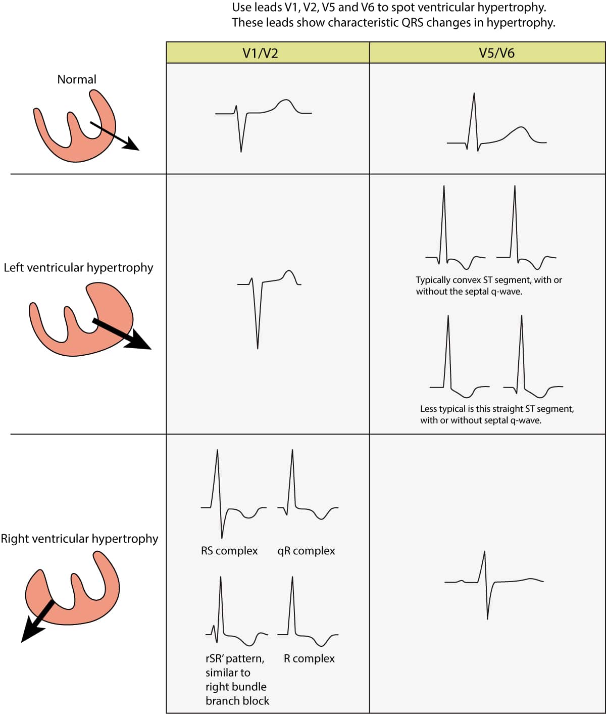
Figure 1. ECG changes seen in left ventricular hypertrophy (LVH) and right ventricular hypertrophy (RVH). The electrical vector of the left ventricle is enhanced in LVH, which results in large R-waves in left sided leads (V5, V6, aVL and I) and deep S-waves in right sided chest leads (V1, V2). Right ventricular hypertrophy causes large R-waves in right sided chest leads and deeper S-waves in left sided leads.