
Figure 1. The ECG contour of the normal P-wave, P mitrale (left atrial enlargement) and P pulmonale (right atrial enlargement)
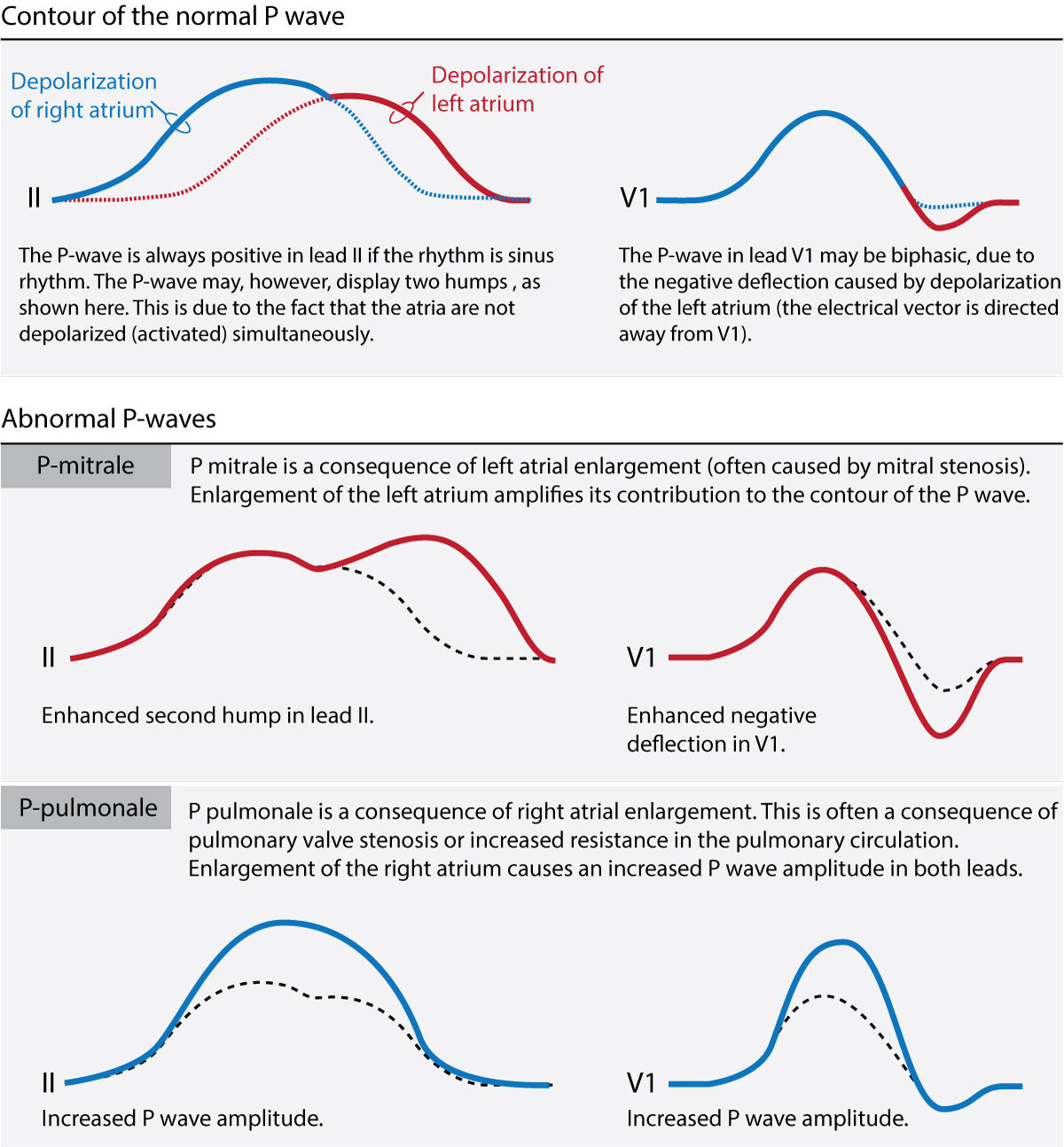


Figure 1. The ECG contour of the normal P-wave, P mitrale (left atrial enlargement) and P pulmonale (right atrial enlargement)