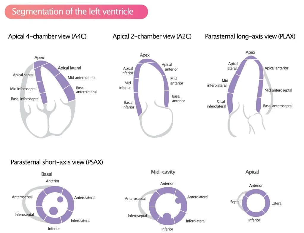
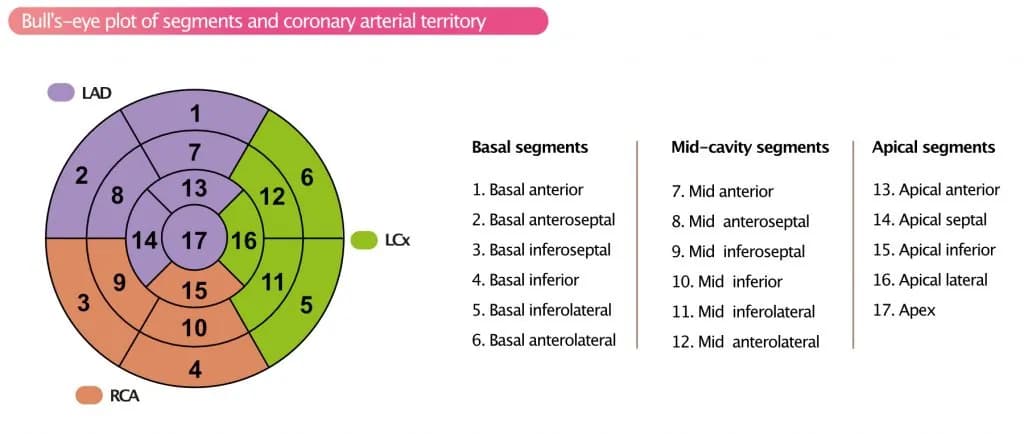
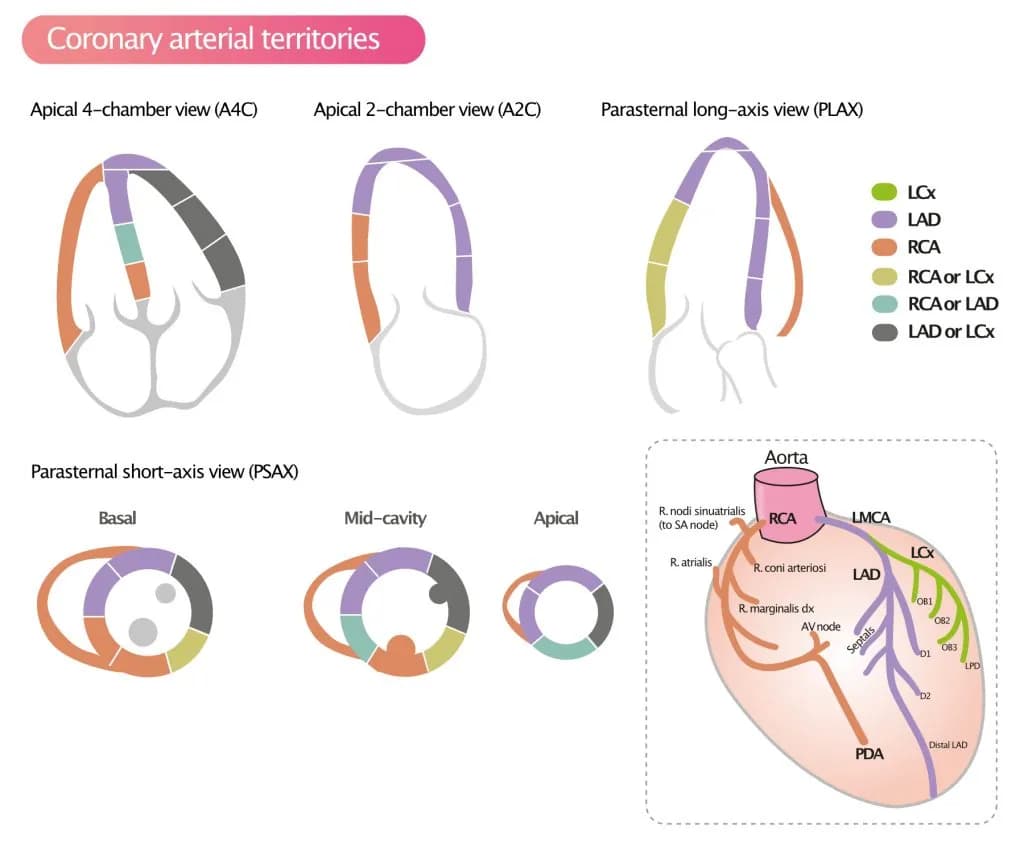
Reference (normal) values for echocardiography
Aorta
Aorta
Normal values for aorta in 2D echocardiography
| Normal interval | Normal interval, adjusted | |
| Aortic annulus | 20-31 mm | 12-14 mm/m2 |
| Sinus valsalva | 29-45 mm | 15-20 mm/m2 |
| Sinotubular junction | 22-36 mm | 13-17 mm/m2 |
| Ascending aorta | 22-36 mm | 13-17 mm/m2 |
| Aortic arch | 22-36 mm | |
| Descending aorta | 20-30 mm |
Adjustment refers to adjusting for body surface area (BSA).
Aorta, predicted
Aorta, standardized
Standardized values for aorta (mean ± SD)
| Aortic annulus | Sinus valsalva | |||
| Age | Women | Men | Women | Men |
| <30 | 11.4 ± 1.1 | 11.4 ± 1.2 | 15.5 ± 1.5 | 15.3 ± 1.8 |
| 30–39 | 11.7 ± 1.0 | 11.4 ± 1.1 | 16.5 ± 1.6 | 15.7 ± 1.3* |
| 40–49 | 11.7 ± 1.0 | 11.5 ± 0.9 | 16.8 ± 1.4 | 16.6 ± 1.5 |
| 50–59 | 12.0 ± 0.9 | 11.5 ± 0.9 | 17.9 ± 1.6 | 16.8 ± 1.7* |
| 60–69 | 11.7 ± 1.1 | 12.1 ± 1.0 | 17.5 ± 1.7 | 17.8 ± 1.6 |
| >70 | 11.6 ± 1.1 | 12.0 ± 1.0 | 18.2 ± 1.9 | 18.1 ± 1.7 |
| All | 11.7 ± 1.0 | 11.6 ± 1.0 | 17.0 ± 1.9 | 16.6 ± 1.8 |
| Sinotubular junction | Ascending aorta | |||
| Age | Women | Men | Women | Men |
| <30 | 13.5 ± 1.4 | 12.7 ± 2.3 | 15.1 ± 1.6 | 14.3 ± 1.6* |
| 30–39 | 14.3 ± 1.5 | 13.3 ± 1.3 | 16.4 ± 1.8 | 14.5 ± 1.4* |
| 40–49 | 14.6 ± 1.2 | 14.1 ± 1.5* | 16.4 ± 1.4 | 15.5 ± 1.5* |
| 50–59 | 15.7 ± 1.3 | 14.1 ± 1.5 | 18.0 ± 1.4 | 16.2 ± 1.6* |
| 60–69 | 14.7 ± 1.6 | 15.1 ± 1.6* | 17.6 ± 1.7 | 17.3 ± 1.7 |
| >70 | 15.5 ± 2.0 | 14.7 ± 1.6 | 18.2 ± 3.3 | 17.5 ± 1.4 |
| All | 14.7 ± 1.7 | 13.9 ± 1.8 | 16.9 ± 2.3 | 15.7 ± 1.8 |
| Aortic arch | ||
| Age | Women | Men |
| <30 | 10.7 ± 1.0 | 10.1 ± 1.3* |
| 30–39 | 11.6 ± 1.6 | 10.8 ± 1.3* |
| 40–49 | 11.5 ± 1.2 | 11.3 ± 1.3 |
| 50–59 | 12.7 ± 1.1 | 11.3 ± 1.2* |
| 60–69 | 12.5 ± 1.4 | 12.0 ± 1.1 |
| >70 | 12.7 ± 1.2 | 12.2 ± 1.3 |
| All | 11.9 ± 1.5 | 11.2 ± 1.4 |
Aortic valve regurgitation
Aortic valve regurgitation, normal values
Primary parameters
| Mild regurgitation | Moderate regurgitation | Severe regurgitation | |
| Vena contracta (cm) | < 0,3 | 0,3-0,6 | > 0,6 |
| PHT (pressure half time) – CW doppler | > 500 | 200-500 | < 200 |
| Diastolic flow reversal – PW doppler aorta descendens | if present, early diastolic | early diastolic | holodiastolic |
Quantification of jet
| Mild regurgitation | Moderate regurgitation | Severe regurgitation | |
| Jet-width / LVOT-width | < 0,25 | 0,25-0,64 | > 0,64 |
| Jet-area / LVOT-area | < 0,05 | 0,05-0,59 | > 0,59 |
Qualitative parameters
| Mild regurgitation | Moderate regurgitation | Severe regurgitation | |
| Jet area | small | variable | large if central, variable if excentric |
| Coaptation defect | mild | moderate | severe |
| CW-profile aortic regurgitation | weak | variable | dense |
| Size left atrium | normal | normal or enlarged | enlarged |
| Size left ventricle | normal | normal or enlarged | enlarged |
Additional PISA-parameters
| Mild regurgitation | Moderate regurgitation | Severe regurgitation | |
| EROA (cm²) | < 0,10 | 0,10-0,29 | > 0,29 |
| Regurgitation volume (ml/beat) | < 30 | 30-59 | > 59 |
| Regurgitation fraction (%) | < 30 | 30-49 | > 49 |
EROA = (CSA*v)/v(maxMR) {{ Flödeshastighet = CSA*v = 6.28*r2*v(aliasing)
References
1. Zoghbi, William A.; Enriquez-Sarano, Maurice; Foster, Elyse; Grayburn, Paul A.; Kraft, Carol D.; Levine, Robert A. et al. (2003): Recommendations for evaluation of the severity of native valvular regurgitation with two-dimensional and Doppler echocardiography. In Journal of the American Society of Echocardiography : official publication of the American Society of Echocardiography 16 (7), pp. 777–802. DOI: 10.1016/S0894-7317(03)00335-3.
2. Zoghbi, William A.; Adams, David; Bonow, Robert O.; Enriquez-Sarano, Maurice; Foster, Elyse; Grayburn, Paul A. et al. (2017): Recommendations for Noninvasive Evaluation of Native Valvular Regurgitation. A Report from the American Society of Echocardiography Developed in Collaboration with the Society for Cardiovascular Magnetic Resonance. In Journal of the American Society of Echocardiography : official publication of the American Society of Echocardiography 30 (4), pp. 303–371. DOI: 10.1016/j.echo.2017.01.007.
Aortic valve stenosis
Aortic valve stenosis
Disease defining parameters
| Mild stenosis | Moderate stenosis | Severe stenosis | |
| Valve area (cm²) | > 1,5 | 1,0-1,5 | < 1,0 |
| Normalized valve area (cm²/m²) | >0,85 | 0,6-0,85 | < 0,6 |
Measured parameters
| Aortic sclerosis | Mild stenosis | Moderate stenosis | Severe stenosis | |
| Mean gradient (mmHg) | < 20 | 20-40 | > 40 | |
| Peak velocity (m/s) | 1,8-2,5 | 2,6-2,9 | 3,0-4,0 | > 4,0 |
| Peak gradient (mmHg) | < 35 | 35-65 | > 65 | |
| Velocity ratio (LVOT / aortic valve) | > 0,5 | 0,5-0,25 | < 0,25 |
References
1. Baumgartner, Helmut; Hung, Judy; Bermejo, Javier; Chambers, John B.; Evangelista, Arturo; Griffin, Brian P. et al. (2009): Echocardiographic assessment of valve stenosis: EAE/ASE recommendations for clinical practice. In Journal of the American Society of Echocardiography : official publication of the American Society of Echocardiography 22 (1), 1-23; quiz 101-2. DOI: 10.1016/j.echo.2008.11.029.
2. Baumgartner, Helmut; Hung, Judy; Bermejo, Javier; Chambers, John B.; Evangelista, Arturo; Griffin, Brian P. et al. (2009): Echocardiographic assessment of valve stenosis: EAE/ASE recommendations for clinical practice. In European journal of echocardiography : the journal of the Working Group on Echocardiography of the European Society of Cardiology 10 (1), pp. 1–25. DOI: 10.1093/ejechocard/jen303.
3. Vahanian, Alec; Alfieri, Ottavio; Andreotti, Felicita; Antunes, Manuel J.; Barón-Esquivias, Gonzalo; Baumgartner, Helmut et al. (2012): Guidelines on the management of valvular heart disease (version 2012). In European heart journal 33 (19), pp. 2451–2496. DOI: 10.1093/eurheartj/ehs109.
4. Baumgartner, Helmut; Hung, Judy; Bermejo, Javier; Chambers, John B.; Edvardsen, Thor; Goldstein, Steven et al. (2017): Recommendations on the Echocardiographic Assessment of Aortic Valve Stenosis. A Focused Update from the European Association of Cardiovascular Imaging and the American Society of Echocardiography. In Journal of the American Society of Echocardiography : official publication of the American Society of Echocardiography 30 (4), pp. 372–392. DOI: 10.1016/j.echo.2017.02.009.
Left and right ventricle
Visual assessment of systolic function
Visual assessment of regional wall motion (left ventricle)
| TYPE OF MOTION | DEFINITION | WALL MOTION SCORE |
| Normal | Normal thickening (typically >30% thickening from end-diastole to end-systole). | 1 |
| Hypokinesia | Reduced thickening (typically 10–30% thickening from end-diastole to end-systole) | 2 |
| Akinesia | Markedly reduced, or no thickening (<10%) | 3 |
| Dyskinesia | Paradoxical thinning and/or outward directed motion during systole | 4 |
| Aneurysmatic | Diastolic deformation | 5 |
Recommended by American Society for Echocardiography (J Am Soc Echocardiogr 18:1440-1463, 2005).
Left ventricular mass and geometry
Reference values for left ventricular mass and geometry
| Women | Men | |||||||
| Reference | Mildy abnormal | Moderately abnormal | Severely abnormal | Reference | Mildly abnormal | Moderately abnormal | Severely abnormal | |
| LINEAR METHOD | ||||||||
| LV mass, g | 67–162 | 163–186 | 187–210 | >211 | 88–224 | 225–258 | 259–292 | >293 |
| LV mass/BSA, g/m2 | 43–95 | 96–108 | 109–121 | > 122 | 49–115 | 116–131 | 132–148 | > 149 |
| LV mass/height, g/m | 41–99 | 100–115 | 116–128 | >129 | 52–126 | 127–144 | 145–162 | >163 |
| LV mass/height 2,7 , g/m2,7 | 18–44 | 45–51 | 52–58 | >59 | 20–48 | 49–55 | 56–63 | >64 |
| Relative wall thickness, cm | 0.22–0.42 | 0.43–0.47 | 0.48–0.52 | >0.53 | 0.24–0.42 | 0.43–0.46 | 0.47–0.51 | >0.52 |
| Septal thickness, cm | 0.6–0.9 | 1.0–1.2 | 1.3–1.5 | > 1.6 | 0.6–1.0 | 1.1–1.3 | 1.4–1.6 | > 1.7 |
| Posterior wall thickness, cm | 0.6–0.9 | 1.0–1.2 | 1.3–1.5 | > 1.6 | 0.6–1.0 | 1.1–1.3 | 1.4–1.6 | > 1.7 |
| 2D METOD | ||||||||
| LV mass, g | 66–150 | 151–171 | 172–182 | > 193 | 96–200 | 201–227 | 228–254 | > 255 |
| LV mass/BSA, g/m2 | 44–88 | 89–100 | 101–112 | > 113 | 50–102 | 103–116 | 117–130 | > 131 |
BSA, Body surface area; LV, left ventricular; 2D, 2-dimensional.
Left ventricular dimension and volume
Referensvärden för vänster kammares storlek
| Women | Men | |||||||
| Normal range | Mildly abnormal | Moderately abnormal | Severely abnormal | Normal range | Mildly abnormal | Moderately abnormal | Severely abnormal | |
| LV DIMENSIONS | ||||||||
| LV diastolic diameter | 3.9–5.3 | 5.4–5.7 | 5.8–6.1 | >6.2 | 4.2–5.9 | 6.0–6.3 | 6.4–6.8 | >6.9 |
| LV diastolic diameter/BSA, cm/m 2 | 2.4–3.2 | 3.3–3.4 | 3.5–3.7 | >3.8 | 2.2–3.1 | 3.2–3.4 | 3.5–3.6 | >3.7 |
| LV diastolic diameter/height, cm/m | 2.5–3.2 | 3.3–3.4 | 3.5–3.6 | >3.7 | 2.4–3.3 | 3.4–3.5 | 3.6–3.7 | >3.8 |
| LV VOLUME | ||||||||
| LV diastolic volume, mL | 56–104 | 105–117 | 118–130 | >131 | 67–155 | 156–178 | 179–201 | >201 |
| LV diastolic volume/BSA, mL/m 2 | 35–75 | 76–86 | 87–96 | > 97 | 35–75 | 76–86 | 87–96 | > 97 |
| LV systolic volume, mL | 19–49 | 50–59 | 60–69 | >70 | 22–58 | 59–70 | 71–82 | >83 |
| LV systolic volume/BSA, mL/m 2 | 12–30 | 31–36 | 37–42 | > 43 | 12–30 | 31–36 | 37–42 | > 43 |
BSA, body surface area; LV, left ventricular.
Left ventricular function (ejection fraction)
Reference values for left ventricular function (ejection fraction)
| Women | Men | |||||||
| Normal | Mildly abnormal | Moderately abnormal | Severely abnormal | Normal | Mildly abnormal | Moderately abnormal | Severely abnormal | |
| LINEAR METHOD | ||||||||
| Endocardial fractional shortening, % | 27–45 | 22–26 | 17–21 | <16 | 25–43 | 20–24 | 15–19 | <14 |
| Midwall fractional shortening, % | 15–23 | 13–14 | 11–12 | <10 | 14–22 | 12–13 | 10–11 | <10 |
| 2D METOD | ||||||||
| Ejection fraction, % | >55 | 45–54 | 30–44 | <30 | >55 | 45–54 | 30–44 | <30 |
2D, Two-dimensional.
Classification of ejection fraction
| Normal | Mildly abnormal | Moderately abnormal | Severely abnormal | |
| Ejection fraction (%), biplan, males | 52-72 | 41-51 | 30-40 | <30 |
| Ejection fraction (%), biplan, females | 54-74 | 41-53 | 30-40 | <30 |
References
1. Lang, Roberto M.; Badano, Luigi P.; Mor-Avi, Victor; Afilalo, Jonathan; Armstrong, Anderson; Ernande, Laura et al. (2015): Recommendations for cardiac chamber quantification by echocardiography in adults: an update from the American Society of Echocardiography and the European Association of Cardiovascular Imaging. In European heart journal cardiovascular Imaging 16 (3), pp. 233–270. DOI: 10.1093/ehjci/jev014.
2. Lang, Roberto M.; Bierig, Michelle; Devereux, Richard B.; Flachskampf, Frank A.; Foster, Elyse; Pellikka, Patricia A. et al. (2005): Recommendations for chamber quantification: a report from the American Society of Echocardiography’s Guidelines and Standards Committee and the Chamber Quantification Writing Group, developed in conjunction with the European Association of Echocardiography, a branch of the European Society of Cardiology. In Journal of the American Society of Echocardiography : official publication of the American Society of Echocardiography 18 (12), pp. 1440–1463. DOI: 10.1016/j.echo.2005.10.005. –>Pubmed-Link
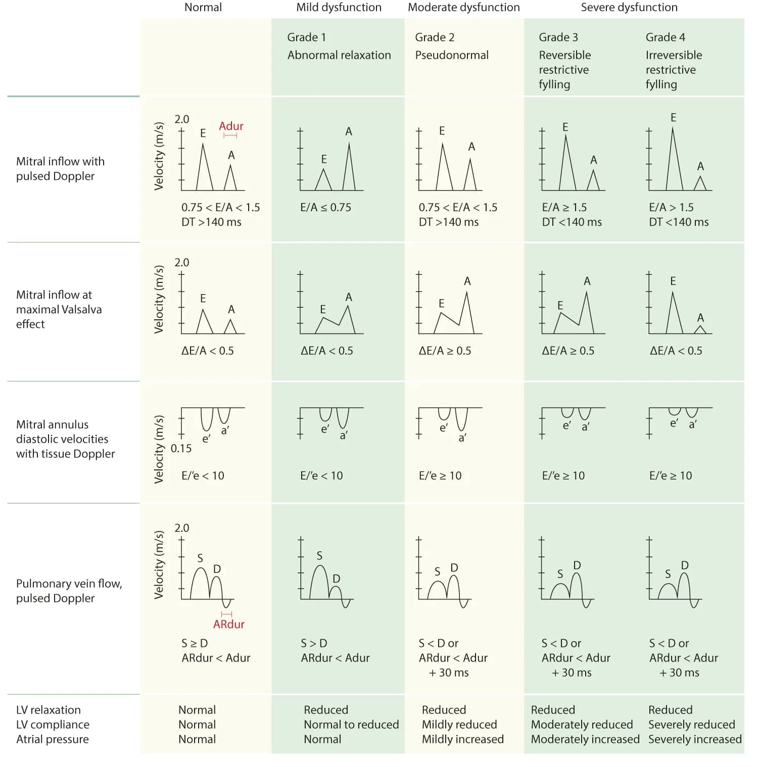
Diastolic function
| Criteria | Normal, young | Normal, adult | Abnormal relaxation (grade 1 dysfunction) | Pseudonormal (grade 2 dysfunction) | Reversible restrictive filling (grade 3 dysfunction) | Irreversible restrictive filling (grade 4 dysfunction) |
| E/A-ratio | 1–2 | 1–2 | <1 | 1–1.5 (not reversed with Valsalva maneuver) | >1.5 | 1.5–2.0 (not reversed with Valsalva maneuver) |
| Deceleration time (DT), ms | <240 | 150–240 | ≥240 | 150–200 | <150 | <150 |
| IVRT (ms) | 70–90 | 70–90 | >90 | <90 | <70 | <70 |
| S/D-ratio | <1 | ≥1 | ≥1 | <1 | <1 | <1 |
| ARdur – Adur (ms) | ≥30 | ≤0 | ≤0 or ≥30 | ≥30 | ≥30 | ≥30 |
| AR speed (cm/s) | <35 | <35 | <35 | ≥35 | ≥35 | ≥35 |
| e’ speed (cm/s) | >10 | >8 | <8 | <8 | <8 | <8 |
Right ventricle & pulmonary artery
| Normal interval | Mildly abnormal | Moderately abnormal | Severely abnormal | |
| RV DIMENSIONS | ||||
| Basal RV diameter (RVD 1), cm | 2.0–2.8 | 2.9–3.3 | 3.4–3.8 | >3.9 |
| Mid RV diameter (RVD 2), cm | 2.7–3.3 | 3.4–3.7 | 3.8–4.1 | >4.2 |
| Base-to-apex length (RVD 3), cm | 7.1–7.9 | 8.0–8.5 | 8.6–9.1 | >9.2 |
| RVOT DIAMETER | ||||
| Above aortic valve (RVOT 1), cm | 2.5–2.9 | 3.0–3.2 | 3.3–3.5 | >3.6 |
| Above pulmonic valve (RVOT 2), cm | 1.7–2.3 | 2.4–2.7 | 2.8–3.1 | >3.2 |
| PA DIAMETER | ||||
| Below pulmonic valve (PA 1), cm | 1.5–2.1 | 2.2–2.5 | 2.6–2.9 | >3.0 |
| RV SIZE AND FUNCTION (MEASURED IN A4C) | ||||
| RV diastolic area, cm2 | 11–28 | 29–32 | 33–37 | >38 |
| RV systolic area, cm2 | 7.5–16 | 17–19 | 20–22 | >23 |
| RV fractional area change, % | 32–60 | 25–31 | 18–24 | <17 |
Left and right atrium
Left and right atrium
Reference values for atrial size and volume
| Women | Men | |||||||
| Normal range | Mildly abnormal | Moderately abnormal | Severely abnormal | Normal range | Mildly abnormal | Moderately abnormal | Severely abnormal | |
| ATRIAL DIMENSIONS | ||||||||
| LA diameter, cm | 2.7–3.8 | 3.9–4.2 | 4.3–4.6 | >4.7 | 3.0–4.0 | 4.1–4.6 | 4.7–5.2 | >5.2 |
| LA diameter/BSA, cm/m2 | 1.5–2.3 | 2.4–2.6 | 2.7–2.9 | >3.0 | 1.5–2.3 | 2.4–2.6 | 2.7–2.9 | >3.0 |
| RA minor-axis dimension, cm | 2.9–4.5 | 4.6–4.9 | 5.0–5.4 | >5.5 | 2.9–4.5 | 4.6–4.9 | 5.0–5.4 | >5.5 |
| RA minor-axis dimension/BSA, cm/m2 | 1.7–2.5 | 2.6–2.8 | 2.9–3.1 | >3.2 | 1.7–2.5 | 2.6–2.8 | 2.9–3.1 | >3.2 |
| ATRIAL AREA | ||||||||
| LA area, cm2 | <20 | 20–30 | 30–40 | >40 | <20 | 20–30 | 30–40 | >40 |
| ATRIAL VOLUMES | ||||||||
| LA volume, mL | 22–52 | 53–62 | 63–72 | >73 | 18–58 | 59–68 | 69–78 | >79 |
| LA volume/BSA, mL/m2 | 22 ± 6 | 29–33 | 34–39 | >40 | 22 ± 6 | 29–33 | 34–39 | >40 |
BSA, Body surface area; LA, left atrial; RA, right atrial.
Mitral valve
Mitral valve regurgitation
Mitral valve regurgitation
Primary parameters
| Mild regurgiation | Moderate regurgitation | Severe regurgitation | |
| Vena contracta (cm) | < 0,3 | 0,3-0,69 | > 0,69 |
| EROA (cm²) | < 0,20 | 0,20-0,39 | > 0,39 |
| Jet size | < 4cm² or < 20% of atrial size | 4-10 cm² or 20-40% of atrial size | > 10 cm² or > 40% of atrial size |
| Jet orientation | central | variable | excentric |
| Pulmonary vein flow | S-dominant | reduced S-wave | systolic flow reversal, |
Qualitative parameters
| Mild regurgiation | Moderate regurgitation | Severe regurgitation | |
| Mitral valve apparatus | normal or mildly abnormal | often abnormal | abnormal, possibly flail |
| CW-profile mitral regurgitation | weak | variable | dense |
| Shape CW-profile mitral regurgitation | parabolic | variable | early peak, triangular |
| Size left atrium | normal | normal or dilated | dilated |
| Size left ventricle | normal | normal or dilated | dilated |
Additional PISA-parameters
| Mild regurgiation | Moderate regurgitation | Severe regurgitation | |
| Regurgitation volume (ml/beat) | < 30 | 30-59 | > 59 |
| Regurgitation volume = EROA x TVI | |||
| Regurgitation fraction (%) | < 30 | 30-49 | > 49 |
Miscellaneous parameters
| Mild regurgiation | Moderate regurgitation | Severe regurgitation | |
| PW-Doppler mitral valve | A dominant | variable | high E values |
References
1. Zoghbi, William A.; Enriquez-Sarano, Maurice; Foster, Elyse; Grayburn, Paul A.; Kraft, Carol D.; Levine, Robert A. et al. (2003): Recommendations for evaluation of the severity of native valvular regurgitation with two-dimensional and Doppler echocardiography. In Journal of the American Society of Echocardiography : official publication of the American Society of Echocardiography 16 (7), pp. 777–802. DOI: 10.1016/S0894-7317(03)00335-3.
2. Zoghbi, William A.; Adams, David; Bonow, Robert O.; Enriquez-Sarano, Maurice; Foster, Elyse; Grayburn, Paul A. et al. (2017): Recommendations for Noninvasive Evaluation of Native Valvular Regurgitation. A Report from the American Society of Echocardiography Developed in Collaboration with the Society for Cardiovascular Magnetic Resonance. In Journal of the American Society of Echocardiography : official publication of the American Society of Echocardiography 30 (4), pp. 303–371. DOI: 10.1016/j.echo.2017.01.007. –>Pubmed-Link
Mitral valve stenosis
Mitral valve stenosis
Primary parameters
| Mild stenosis | Moderate stenosis | Severe stenosis | |
| Valve area | > 1,5 cm² | 1,5 – 1,0 cm² | < 1,0 cm² |
| Mean gradient | < 5 mmHg | 5-10 mmHg | > 10 mmHg |
Additional parameters
| Mild stenosis | Moderate stenosis | Severe stenosis | |
| Pulmonary artery pressure | <30 | 30-50 | >50 |
References
1. Baumgartner, Helmut; Hung, Judy; Bermejo, Javier; Chambers, John B.; Evangelista, Arturo; Griffin, Brian P. et al. (2009): Echocardiographic assessment of valve stenosis: EAE/ASE recommendations for clinical practice. In European journal of echocardiography : the journal of the Working Group on Echocardiography of the European Society of Cardiology 10 (1), pp. 1–25. DOI: 10.1093/ejechocard/jen303.
2. Baumgartner, Helmut; Hung, Judy; Bermejo, Javier; Chambers, John B.; Evangelista, Arturo; Griffin, Brian P. et al. (2009): Echocardiographic assessment of valve stenosis: EAE/ASE recommendations for clinical practice. In Journal of the American Society of Echocardiography : official publication of the American Society of Echocardiography 22 (1), 1-23; quiz 101-2. DOI: 10.1016/j.echo.2008.11.029.
3. Vahanian, Alec; Alfieri, Ottavio; Andreotti, Felicita; Antunes, Manuel J.; Barón-Esquivias, Gonzalo; Baumgartner, Helmut et al. (2012): Guidelines on the management of valvular heart disease (version 2012). In European heart journal 33 (19), pp. 2451–2496. DOI: 10.1093/eurheartj/ehs109.
Tricuspid valve
Tricuspid valve regurgitation
Tricuspid valve regurgitation
Primary parameters
| Mild regurgiation | Moderate regurgitation | Severe regurgitation | |
| PISA radius | < 0,6 | 0,6-0,9 | > 0,9 |
| Vena contracta (cm) | not defined | < 0,7 | > 0,7 |
| Liver vein flow | S-dominant | Reduced S-wave | systolic flow reversal, jet reaching liver veins |
Additional quantitative parameters
| Mild regurgiation | Moderate regurgitation | Severe regurgitation | |
| Jet size | < 5cm² | 5-10 cm² | > 10 cm² |
| size vena cava inferior | < 2,1 cm and compressible | in between | > 2,1 cm, almost not compressible |
Additional qualitative parameters
| Mild regurgiation | Moderate regurgitation | Severe regurgitation | |
| Coaptation defect | mild | moderate | severe |
| Size right ventricle | normal | normal or enlarged | enlarged |
| Size right atrium | normal | normal or enlarged | enlarged |
| CW-profile tricuspid regurgitation | weak | variable | dense |
| Shape CW-profile tricuspid regurgitation | parabolic | variable | early peak, triangular |
References
1. Zoghbi, William A.; Enriquez-Sarano, Maurice; Foster, Elyse; Grayburn, Paul A.; Kraft, Carol D.; Levine, Robert A. et al. (2003): Recommendations for evaluation of the severity of native valvular regurgitation with two-dimensional and Doppler echocardiography. In Journal of the American Society of Echocardiography : official publication of the American Society of Echocardiography 16 (7), pp. 777–802. DOI: 10.1016/S0894-7317(03)00335-3.
2. Zoghbi, William A.; Adams, David; Bonow, Robert O.; Enriquez-Sarano, Maurice; Foster, Elyse; Grayburn, Paul A. et al. (2017): Recommendations for Noninvasive Evaluation of Native Valvular Regurgitation. A Report from the American Society of Echocardiography Developed in Collaboration with the Society for Cardiovascular Magnetic Resonance. In Journal of the American Society of Echocardiography : official publication of the American Society of Echocardiography 30 (4), pp. 303–371. DOI: 10.1016/j.echo.2017.01.007. –>Pubmed-Link
Tricuspid valve stenosis
Tricuspid valve stenosis
| Mean pressure gradient | ≥ 5 mmHg |
| Valve area according to the continuity equation (cm²) | ≤ 1 cm² |
| ZTime-velocity-integral of the tricuspid inflow (cm) | > 60 cm |
| Pressure half time (T ½,ms) | ≥ 190 ms |
| Right atrium | Severly enlarged |
| Vena cava inferior | enlarged |
References
1. Baumgartner, Helmut; Hung, Judy; Bermejo, Javier; Chambers, John B.; Evangelista, Arturo; Griffin, Brian P. et al. (2009): Echocardiographic assessment of valve stenosis: EAE/ASE recommendations for clinical practice. In European journal of echocardiography : the journal of the Working Group on Echocardiography of the European Society of Cardiology 10 (1), pp. 1–25. DOI: 10.1093/ejechocard/jen303.
2. Baumgartner, Helmut; Hung, Judy; Bermejo, Javier; Chambers, John B.; Evangelista, Arturo; Griffin, Brian P. et al. (2009): Echocardiographic assessment of valve stenosis: EAE/ASE recommendations for clinical practice. In Journal of the American Society of Echocardiography : official publication of the American Society of Echocardiography 22 (1), 1-23; quiz 101-2. DOI: 10.1016/j.echo.2008.11.029. –>Pubmed-Link
Pulmonary valve
Pulmonary valve regurgitation
Pulmonary valve regurgitation
| Mild regurgiation | Moderate regurgitation | Severe regurgitation | |
| Pulmonary valve morphology | normal | normal/abnormal | abnormal |
| Density CW-Doppler | weak, slow deceleration | variable | dense, steep deceleration, early termination of diastolic flow |
| Jet width colour Doppler | small,, < 10 mm length, narrow origin | variable | large, wide origin |
| Ratio pulmonic to aortic flow | normal | variable | significantly increased |
References
1. Zoghbi, William A.; Enriquez-Sarano, Maurice; Foster, Elyse; Grayburn, Paul A.; Kraft, Carol D.; Levine, Robert A. et al. (2003): Recommendations for evaluation of the severity of native valvular regurgitation with two-dimensional and Doppler echocardiography. In Journal of the American Society of Echocardiography : official publication of the American Society of Echocardiography 16 (7), pp. 777–802. DOI: 10.1016/S0894-7317(03)00335-3.
2. Zoghbi, William A.; Adams, David; Bonow, Robert O.; Enriquez-Sarano, Maurice; Foster, Elyse; Grayburn, Paul A. et al. (2017): Recommendations for Noninvasive Evaluation of Native Valvular Regurgitation. A Report from the American Society of Echocardiography Developed in Collaboration with the Society for Cardiovascular Magnetic Resonance. In Journal of the American Society of Echocardiography : official publication of the American Society of Echocardiography 30 (4), pp. 303–371. DOI: 10.1016/j.echo.2017.01.007.
3. Lancellotti, Patrizio; Tribouilloy, Christophe; Hagendorff, Andreas; Moura, Luis; Popescu, Bogdan A.; Agricola, Eustachio et al. (2010): European Association of Echocardiography recommendations for the assessment of valvular regurgitation. Part 1: aortic and pulmonary regurgitation (native valve disease). In European journal of echocardiography : the journal of the Working Group on Echocardiography of the European Society of Cardiology 11 (3), pp. 223–244. DOI: 10.1093/ejechocard/jeq030.
Pulmonary valve stenosis
Pulmonary valve stenosis
| Mild stenosis | Moderate stenosis | Severe stenosis | |
| Peak velocity (m/s) | < 3 | 43528 | > 4 |
| Peak gradient (mmHg) | < 36 | 36-64 | > 64 |
References
1. Baumgartner, Helmut; Hung, Judy; Bermejo, Javier; Chambers, John B.; Evangelista, Arturo; Griffin, Brian P. et al. (2009): Echocardiographic assessment of valve stenosis: EAE/ASE recommendations for clinical practice. In European journal of echocardiography : the journal of the Working Group on Echocardiography of the European Society of Cardiology 10 (1), pp. 1–25. DOI: 10.1093/ejechocard/jen303.
2. Baumgartner, Helmut; Hung, Judy; Bermejo, Javier; Chambers, John B.; Evangelista, Arturo; Griffin, Brian P. et al. (2009): Echocardiographic assessment of valve stenosis: EAE/ASE recommendations for clinical practice. In Journal of the American Society of Echocardiography : official publication of the American Society of Echocardiography 22 (1), 1-23; quiz 101-2. DOI: 10.1016/j.echo.2008.11.029. –>Pubmed-Link